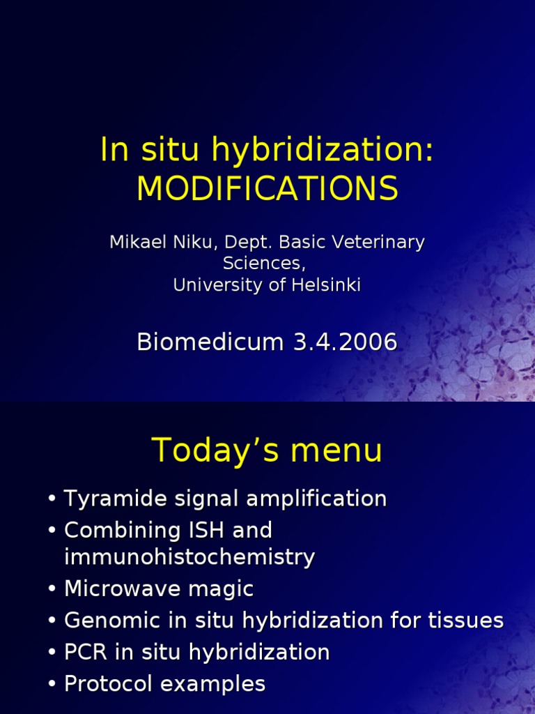
In situ hybridization (ISH) is a pivotal technique employed in molecular biology and histology that facilitates the localization of specific nucleic acid sequences within fixed tissues and cells. It is a powerful method that provides insights into the spatial and temporal expression of genes, thereby unveiling the intricate tapestry of biological systems. At its core, in situ hybridization serves as a bridge between the molecular realm and the broader organismal context, rendering it an indispensable tool in the arsenal of researchers and clinicians alike.
The essence of in situ hybridization lies in its ability to detect RNA or DNA within the cellular or tissue milieu. This technique involves the use of labeled probes that are complementary to the target nucleic acid sequence. These probes hybridize to the specific regions of interest, allowing researchers to observe the distribution and abundance of genetic material within the cellular architecture. One might wonder why such an approach is met with widespread fascination among scientists. The answer lies not merely in its technical procedure but in the wealth of information it unveils about gene expression, tissue development, and the diagnosis of diseases.
One primary observation in the application of in situ hybridization is the striking spatial heterogeneity often seen in gene expression. Different cells or regions of a tissue can display stark variations in the presence or absence of certain transcripts, suggesting a complexity that calls for deeper investigation. This differential expression is not merely a trivial detail; rather, it holds significant implications for understanding developmental processes, disease states, and responses to environmental cues. For example, in the context of embryonic development, spatially regulated gene expression orchestrates the intricate process of differentiation, leading to the formation of diverse cell types and tissues.
The implementation of in situ hybridization provides clarity on how specific genes govern particular biological pathways. Consider the study of cancer, where aberrant gene expression often correlates with tumor progression and metastasis. Through in situ hybridization, researchers can pinpoint the exact locations of oncogenes and tumor suppressor genes in a tumor sample. Such precise localization reveals not only the presence of these genes but can also indicate the phase of malignancy and potential prognostic outcomes. Hence, the technique transcends mere elucidation; it empowers prognostic and therapeutic decisions that could significantly alter patient management.
The methodology behind in situ hybridization has evolved considerably since its inception. Initially, the technique relied on radioactive probes, which, while effective, posed safety concerns and practical limitations. The advent of non-radioactive, fluorescent, or chromogenic probes has revolutionized the approach, yielding images that are more accessible and safer to produce. Fluorescent in situ hybridization (FISH), in particular, has gained prominence due to its ability to visualize multiple targets simultaneously, enhancing the depth of spatial analysis. The multiplexing capabilities of FISH have opened new avenues in genomic research, enabling simultaneous investigations of complex interactions among different genes.
Beyond the laboratory, in situ hybridization holds promise in clinical settings as a diagnostic tool. As we delve into the field of personalized medicine, the ability to accurately assess gene expression profiles within tissues is indispensable. A thorough understanding of the molecular dynamics governing a patient’s unique tumor can dictate tailored therapeutic regimes. For instance, measuring expression levels of receptors or oncogenes can categorize tumors into distinct subtypes, guiding treatment options and potentially improving patient outcomes. This movement toward personalized approaches underscores the necessity of integrating molecular diagnostics into routine clinical practice.
Despite its virtues, in situ hybridization is not without limitations. The inherent complexity of tissues, varying levels of probe accessibility, and potential non-specific binding can yield ambiguous results. Interpretation of the data requires a nuanced understanding of both the biological context and the intricacies of the technique itself. Moreover, advancements in technologies such as next-generation sequencing (NGS) are reshaping the landscape of gene expression analysis, raising questions regarding the future prominence of in situ hybridization in the face of rapid technological evolution.
The fascination with in situ hybridization goes beyond its technical aspects and tangible outputs. It is profoundly rooted in the quest to understand life’s molecular underpinnings amidst a universe of complexity. The application of this technique resonates with the broader narrative of biology, where the synthesis of molecular signals and structural diversity converges to form the tapestry of life. In studying the intricate interplay of genes within their native contexts, we gain invaluable insights not only into the mechanisms of health and disease but also into the very fabric of evolution and adaptation.
In conclusion, in situ hybridization exemplifies the merger of art and science in revealing the profound complexity of biological systems. As researchers continue to develop and refine methodologies surrounding this technique, it is likely that our understanding of gene expression and its implications will deepen. As we navigate through understanding life’s molecular biographies, in situ hybridization remains a beacon that illuminates the pathways through which genes shape organismal function, health, and disease.
